Before the appearance of genetics, the classification of living organisms was made by bringing together their common characteristics. These similarities made it possible to position them in an order reflecting the position they occupy in the evolution tree. The grouping of common characters is done around an entity named taxon. For example, the species is the last taxon within the systematic classification, but if we consider not the species but the family, all species of this family are in turn taxa of this family.
The classification of springtails therefore involves an identification of these common traits, the dissociation of which may require very detailed studies. For example, we can study the size or position of the hair and sensilla, ocelli within the ocular plate, the postantennial organ, various pigmentations, antennas, and other organs or external appendages. In many cases, a binocular or microscope study is necessary. All these elements of identification are called "keys" (examples below) 1
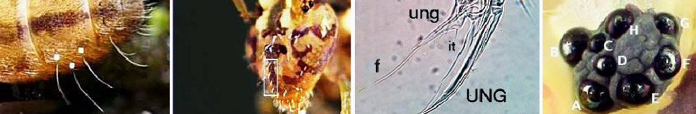
Without proper equipment some elements are not accessible. On the other hand, keys visible on photographs often make it possible to define the belonging to a species or a family. In the example below, a very brief observation of the three springtails (Poduromorphs) can lead to an erroneous identification, whereas if we look more closely specific characters are distinguished.
Identification keys :
Below, I propose to examine some simple keys concerning the identification of the species of three springtail orders. There are few works devoted to this subject, I will however mention the excellent publication resulting from the works of Steve Hopkin: "A key to the Collembola of Britain and Ireland"
Poduromorphes :
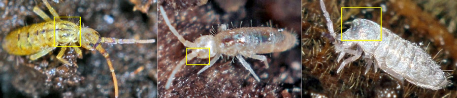
The three specimens observed 1, 2, 3:
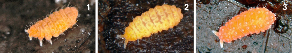
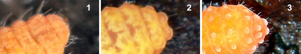
On more detailed images (relating to the 3 specimens above), we can see on the head side: On (1), presence of two ocelli and absence of ocelle visible on (2) and (3). On (1) and (2), no visible tubers and on (3), tubers arranged in order 1 + 2 + 3 + 6.
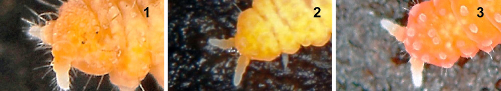
Posterior part: On (1), ending with 1 lobe and on (2) and (3) two lobes.
These simple observable differences from photographs make it possible to identify (1) and (3) respectively as Monobella grassei ("mono" with reference to the end in 1 lobe) and Vitronura gisellae. Springtail (2) can be classified as Neanurinea (body ending in a double lobe).
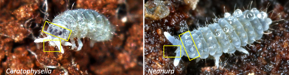
In the Poduromorphs (above), dark and distinct eye plates as well as a wide and rounded buccal cone characterize Ceratophysella (left), while indistinct ocular plaques and a thin, elongated oral cone are distinguished in Neanura (right)
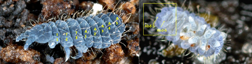
If we look at the last image of Neanura, at first glance, it is not easy to distinguish it from Deutonura (above left). We must then refer to the abdominal segments. In Neanura, the six abdominal segments are visible (we distinguish the last abdominal segment (6) on the previous image, in the form of two juxtaposed lobes), in Deutonura only the fifth segment is visible (A5) and it completely covers the sixth segment .
Symphypleones :
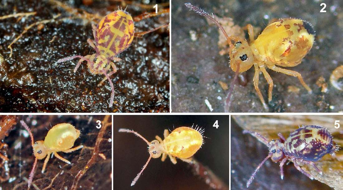
The Symphypleones above have strong similarities. The dissociation elements of these five distinct species are here at the level of pigmentation:
- Dorsal pigmentation:
Presents = Dicyrtomina ornata (1), Dicyrtomina saundersi (5)
Absent = Dicyrtomina minuta. (2)
- Posterior dark spot:
Absente = Dicyrtomina flavosignata (3) Dicyrtomina signata (4) (in the latter, two shades of colors involved: pale yellow - dark yellow)
Homogeneous rectangle = Dicyrtimona Ornata (1),
Rectangular with streaks horizontally = Dicyrtomina saundersi (5).

Concerning the Dicyrtominae, one can note on the image above that a difference exists between male and female at the level of the pigmentation of the cheeks. If it is pale it is a female (left), if it is dark, we have to do to a male (right).
Entomobryomorphes :
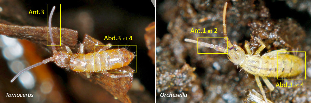
Above, to distinguish between Orchesella (on the right) and Tomocerus (on the left), we must refer to antennas and abdominal segments: In Tomocerus, the third antennal segment is long whereas in Orchesella, first and second segment are subdivided . In Tomocerus, the third abdominal segment and longer than the fourth, while in Orchesella, the third abdominal segment is shorter than the fourth segment (the abdominal segments are positioned from 1 to 6 from the thorax).
Sometimes it is the hair that distinguishes two species. Here (top left) the absence of macrosetae makes it possible to determine in favor of a Desoria, while the presence of macrosetae, on the last abdominal segments, tells us that we are in the presence of an Isotomurus. (Macrosetae are bristles (or hairs) larger than the rest of the setae (bristles or short hairs that cover parts of the body).
Here are three more examples of easily observable details that help in the determination. From left to right:
1) Orchesella villosa, characterized by three dark lines on the first thoracic segment and distinguishing it from other species of Orchesellidae.
2) Heteromurus nitidus, this species is identifiable by the presence of a small red eye.
3) Lepidocyrtus curvicolis, the second thoracic segment (mesothorax) that seems to be the first is very strongly curved (in fact, the first segment or prothorax is reduced and is not visible).
1: First illustration: Montage of 4 images from www.collembola.org. All other montages are made from © Philippe Garcelon.