The cuticle is with the springtails what the skin is to the human, but the comparison stops there because this thin film which covers the whole body of a springtail has astonishing properties. Regarding its constitution, if we refer to the right part of the diagram below1, we see that its structure is fairly standard. It has three layers: the epicuticle (superficial), the exocuticle (intermediate) and a deep layer, the endocuticle. At the level of the epicuticle, the photo (on the left below) taken with an electron microscope shows an arrangement made of a hexagonal assembly of which each element with a size of about one micron is 0.001 mm (other forms exist according to the species).
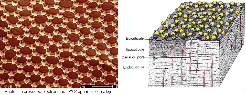
To what necessity can such a disposition, so precisely ordered in perfect geometry and regularity, respond? It can not be the result of chance. The study of the physiological functions of springtail tells us that it breathes through its cuticle and that a desiccation of the latter would be fatal, which is why it likes wetlands. But the excess humidity would be just as fatal if he covered his cuticle. It is therefore mainly to protect against the risk of asphyxiation that springtails has developed a cuticle whose nanostructure has a complex organization that allows it to effectively repel water. In order to allow a good cutaneous respiration, the cuticle must indeed remain clean, dry and free from any stain which could reduce its functional surface. Indeed, the environments in which collembolans evolve are filled with various micro-organisms or water enriched with surfactants (compounds that modify the surface tension resulting from the vegetable decomposition and which, as a result, go against hydrophobic properties intrinsic to the cuticle).
In the images below2, we can observe the same portion of cuticle under three different magnifications. They reveal the granular appearance and fairly homogeneous distribution of small tubers covered with nanotubers. It is precisely the shape of the latter that gives them hydrophobic properties.
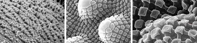
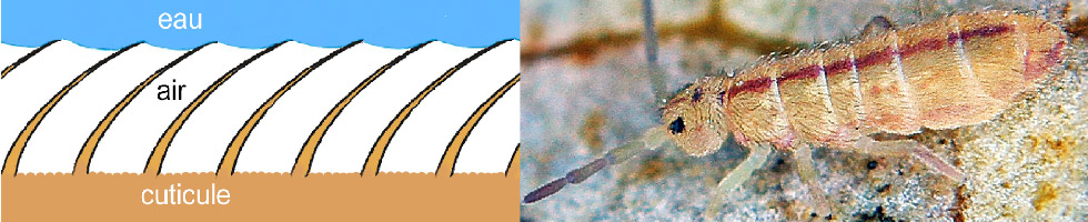
At their contact, the water can not penetrate the interstices which remain in relation with the imprisoned air and allow gas exchanges allowing breathing. This feature is due to the surfactant properties of liquids. The illustration below shows what happens: When a liquid comes into contact with springtail, it also traps air that is confined in a pocket delimited by the interconnect ridges (in white on the diagram on the left) which connect the nano-tubercles to each other. Therefore, the air can continue to provide for the respiratory needs of springtail, even if it is completely submerged.

It should also be noted that the cuticle of most of the springtail is coated with a kind of wax, secreted by specialized glands on the abdomen, which contributes to its surface protection.
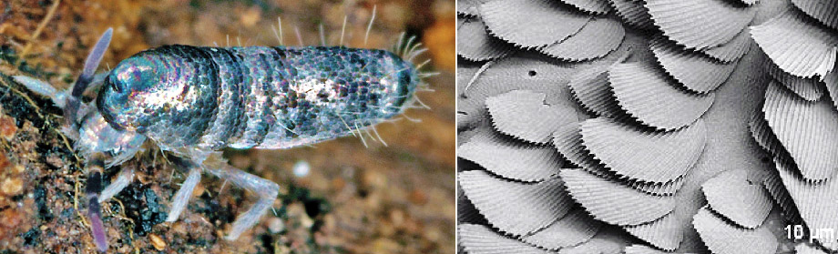
Some springtails, in particular Entomobryidae, have a cuticle similar to other species but which have the particularity of being covered with protective scales. This is for example the case of Lepidocyrtus curvicollis (below) 3-2
Other springtails also have abundant hair that helps to provide them with double protection. Here again, the density of the hairs gives them hydrophobic properties. This is for example the case with isotomurus gramini3 which has a fine hair covering its body homogeneously.
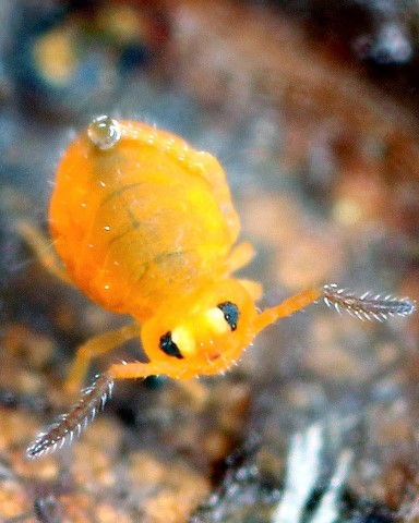
Below is an example4 where the hydrophobic properties of the springtail cuticle are highlighted. Here we must take into account the evaporation of the water contained in the substrate and the increase of the humidity rate of the air which follows. In contact with a cold surface this moisture condenses in the form of fine droplets, as can be seen in nature on various media (herbs, mosses, cobwebs etc ...).

In the case of springtail above (Dicyrtomina ornata3) the thing is not so simple because it is not because of the only condensation that we observe the formation of droplets. Indeed, the structure of the springtail epicuticle forms an open cell pattern (review the sectional view of the cuticle) in which the humidified air is trapped. The absence of air flow within this structure will result in increasing the moisture content which, becoming higher than that of the ambient air, allows the formation of condensation; not because of a difference in temperature but because of the difference in moisture content, because at t ° constant, the dew point (formation of droplets) depends on the saturation differential of the air in water (for understand this phenomenon, see: dew point).
Under certain conditions, it will be possible to observe the formation of these fine droplets due to condensation on the one hand and, on the other hand, the hydrophobic properties of the cuticle (mentioned above). The cuticular pillars, by their structure and also because of their lipid-rich surface, reject these droplets on the surface. The finest of them then agglomerate to form larger and larger liquid spheres until gravity pulls them to the ground.
 Beyond liquids, the cuticle can be soiled by other vegetable or mineral detritus found in nature. Researchers have even tried to deposit fungi or bacteria (Escherichia coli, Staphylococci and Candida) without the latter being able to attach to it. It should be noted that regardless of its intrinsic properties, the cuticle is the subject of careful attention from collembola. The latter using one of his paws comes to take from his mouth a drop of "saliva" with which he will travel the surface of his body including its antennas (see video). This "saliva" from glands called labial nephridia (labial kidneys) could be likened to a kind of urine produced by the latter, located near the mouth. The drop thus produced can also be taken from the collophore or it is transported via the "linea ventralis". As it passes, this drop will play a role of sponge and agglomerate with it any substance or organism that it meets. Thus, the cuticle examined under the microscope, even after being soiled, will soon become clean again and will protect the collembola durably from a large number of external aggressions.
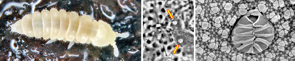
Note: On the skin of some springtails families are disseminated other types of cell structures called pseudocelli. For example, the Poduromorphs, more precisely the Onychiurudae, bear on the head, the thorax and the abdomen. When these springtails are disturbed, their pseudocelli have glands that secrete defensive chemicals. Below is a Kalaphorura burmeisteri3 . The image of the center shows the location of the pseudocelli located at the base of the antennas. The image on the right shows a pseudocelli of Protaphorura4 observed under an electron microscope.
1 Modified sketch, taken from Eisenberg and Richard (1987) in "Biology of the springtails" Stephen P. Hopkin -1997.
2 Photo-cropped from illustrations of "Smart skin patterns protect springtails" - R.Helbig / J.Neckerl / C.Neinhuis / C.Werner- PLOS-30/09/2011.
3 Photos by Philippe Garcelon.
4 According to Frans Janssens, Department of Biology, University of Antwerp collembola.org and "The springtail cuticle ..."
5 Image from a blog by Aron D.Katz - University of Illinois - UIUC- dpt Entomology.